banner image

Ultrasound is a diagnostic technique that generates images of tissues inside the body. The probe generates high-frequency sound waves. Reflected echoes are detected and analyzed to create a real-time, 2-D image.

Ultrasound is non-invasive and is considered to be very safe. Ultrasound is used to diagnose illness, characterize anatomy and guide the direction of diagnostic sampling.

We provide the following outpatient services for cats and dogs at our facility in Mountlake Terrace:

  • Ultrasound
  • Ultrasound-guided centesis
  • Ultrasound-guided fine needle aspirates
  • Ultrasound-guided Tru-Cut biopsy

Pet owners may call our office to schedule an appointment at our facility or submit an inquiry online

image

Frequently Asked Questions

  • General
  • Ultrasound

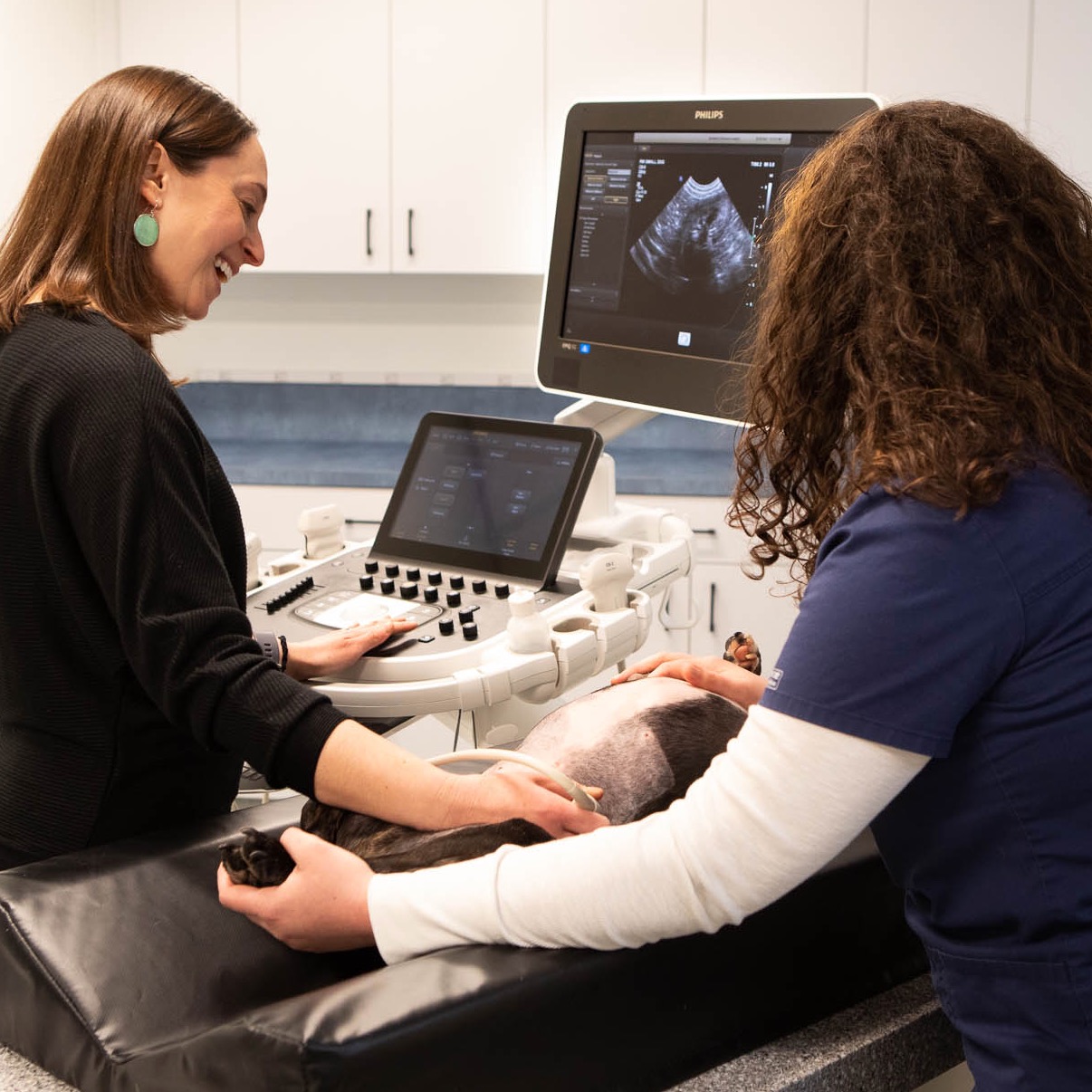
The doctor will provide initial impressions with you that same day.  Complete results will be provided to your veterinarian in the form of a written report within 24 hours.  Your veterinarian will also be provided with access to the images for review.

We can take chest or abdominal radiographs in conjunction with another service like ultrasound. We do not offer outpatient radiographs as a stand-alone service.

The role of fur or hair is to trap air near the skin in order to regulate body temperature. Unfortunately, ultrasound waves cannot pass through air. The fur must be clipped in order to obtain a clear picture.

Most pets require no sedation and lay quietly for the procedure.

Especially nervous or wiggly patients may need sedation to enable a thorough examination.

The structures in the abdomen can be best seen when the stomach is empty. We ask that you withhold food from your pet for 8 hours before your appointment. Water and medications are allowed. Diabetic patients will have special instructions, please contact our staff to discuss fasting.

A thoracic ultrasound is used to assess structures within the chest cavity. We may also evaluate blood flow to the organs within the chest.

An echocardiogram uses live ultrasound images to assess the function and structure of the heart for the specific purpose of assessing heart disease. While we usually briefly evaluate the heart during a thoracic ultrasound, it does not take the place of an echocardiogram.

A fine needle aspirate (FNA) provides a sample of cells taken with a small gauge needle. This procedure may or may not require sedation.

A Tru-Cut biopsy provides a tissue sample requiring a specialized large bore needle. This procedure is performed under general anesthesia to allow for accurate sampling and to reduce the risk of bleeding.  We require a current coagulation profile to ensure that the patient is able to clot appropriately.