Elderly Brussels Griffon
16-year-old MN Brussels Griffon
The patient presented with a two-day history of vomiting, anorexia and lethargy.
On physical examination, the patient was noted to be 8% dehydrated. He was mildly painful and a mass effect was palpated in the mid-abdomen.
CBC: leukocytosis 38.1 K/uL, neutrophilia 34.8 K/uL, lymphopenia 0.47 K/uL, monocytosis 2.5 K/uL
Chemistry: elevated ALT >1000 U/L, ALKP 993 U/L, GGT 129 U/L, t.bili 8.6 mg/dL, BUN 46 mg/dL
cPL: negative
The patient had been treated at the RDVM for 24 hours with IV fluids, Cerenia, Metronidazole and Enrofloxacin. While the vomiting had resolved, the patient was still anorectic and painful. The patient was referred to our facility for an urgent abdominal ultrasound.
Gallbladder Mucocele
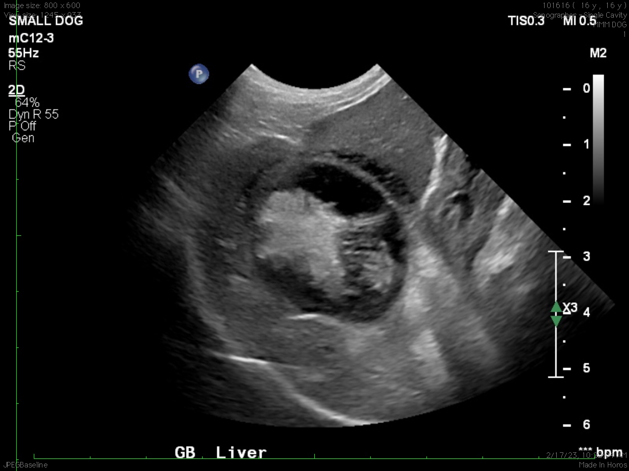
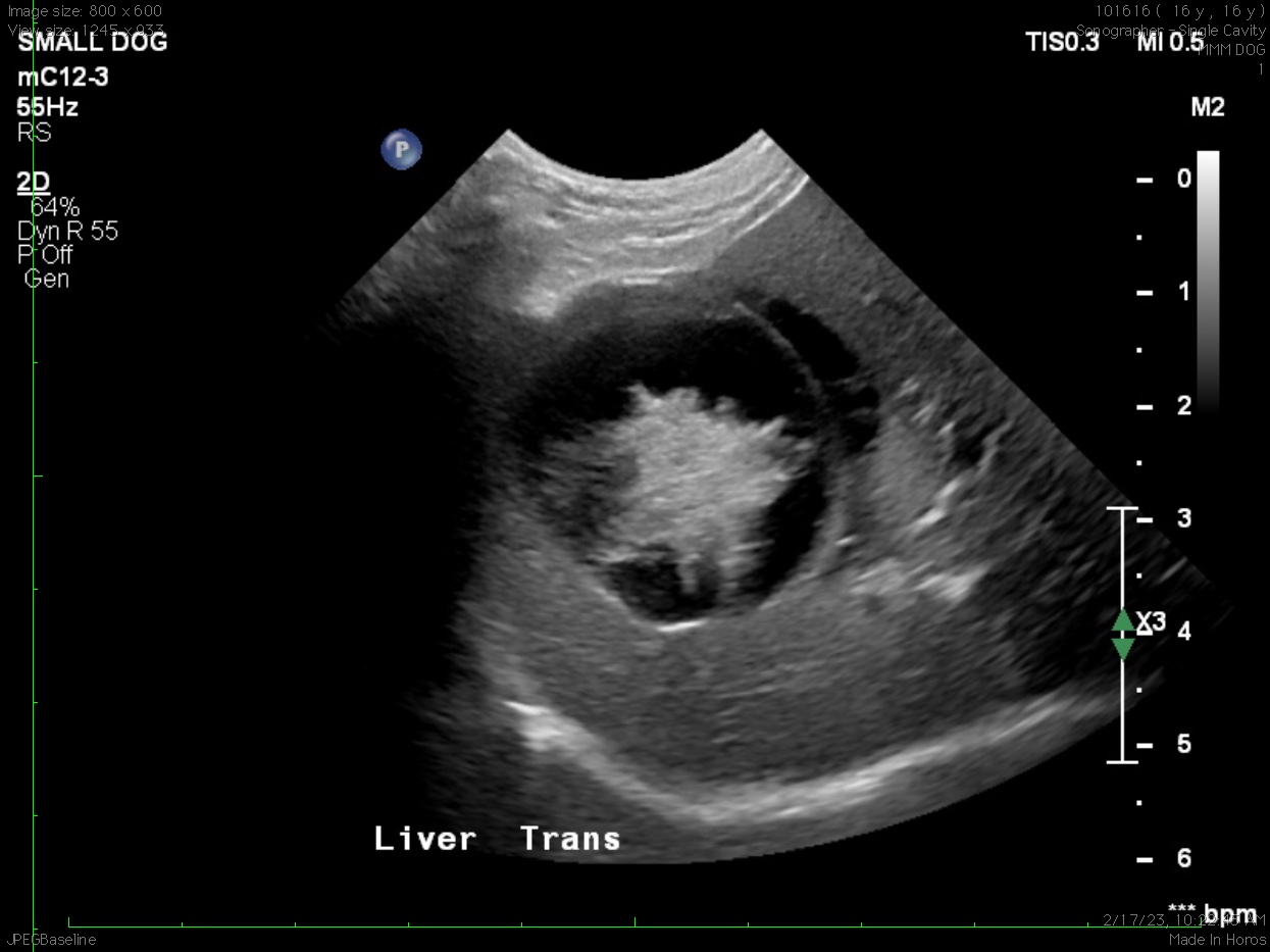
Ultrasound Findings:
The gallbladder is distended, rounded, and has a turgid appearance and contains organized echogenic material in the central lumen. There is marked inflammation of fat as well as a small volume of effusion or edema adjacent to the gallbladder. The liver may be slightly large and has coarse echotexture.
Conclusion:
Gallbladder mucocele
Recommendations:
Ideally, this is a surgical case but if surgery is not an option given the age of the patient then aggressive medical management has reasonable chance at resolving the mucocele. Fluid therapy, pain management, antibiotics, and ursodiol may be necessary. Consider follow-up ultrasound of the gallbladder in 7–10 days.
Discussion:
Gallbladder mucocele (GBM) is an abnormal accumulation of inspissated bile or thick, immobile mucous within the gallbladder lumen. This can lead to gallbladder distention, biliary tract obstruction, and eventually gallbladder necrosis, rupture, and peritonitis. The disease occurs most often in older dogs (mean age of 10 years) with a genetic component being suspected due to strong breed predispositions (e.g. Shetland sheepdog, cocker spaniel, miniature schnauzer). It can also occur in cats, but is rare.
Physical exam and laboratory findings: Clinical signs can range from asymptomatic to very ill due to peritonitis secondary to gallbladder rupture. Clinical signs may include vomiting, inappetence, lethargy, diarrhea, weight loss, abdominal pain, dehydration, icterus, and fever.
Diagnostics: The most significant labwork findings typically include increased serum ALP, ALT, GGT, AST, cholesterol, and bilirubin. We may also see a leukocytosis, neutrophilia, lymphopenia, monocytosis, eosinopenia, thrombocytopenia, and thrombocytosis.
Ultrasonography: Ultrasonography is the best non-invasive method for diagnosis. GBM typically appears as an enlarged gallbladder containing nongravity-dependent contents, with a finely striated or stellate pattern. This finding is similar in appearance to a sliced kiwi. Gallbladder wall thickness is variable. The common bile duct may be dilated. Gallbladder rupture can also be detected with ultrasound. In one study, sensitivity for ultrasonographic identification of gallbladder rupture was 56.1%, and specificity was 91.7%.
Surgical Therapy: Cholecystectomy is the treatment of choice because the risk of future gallbladder rupture is present, even in asymptomatic patients. Complications of surgery include bile peritonitis, sepsis, disseminated intravascular coagulation, and dehiscence.
Medical Therapy: Medical management of GBM is somewhat controversial and is only recommended if no evidence of bile leakage or gallbladder wall necrosis exists. Immediate supportive care may include antiemetics, analgesics, and antimicrobials. Antibiotic therapy is ideally based on culture and sensitivity, but ampicillin and enrofloxacin have been used emperically. Medical therapy may include use of choleretics and hepatoprotectants. Ursodeoxycholic acid (ursodiol) is both a choleretic and hepatoprotectant. Medical therapy is typically continued for a minimum of 4-8 weeks. A recheck abdominal ultrasound is recommended to ensure resolution of the GBM.
Prognosis: Gallbladder mucocele is a potentially life-threatening disease in dogs. In one report, postoperative hypotension and elevations in serum lactate were negatively associated with survival. Another study reported that 17.4% of 219 dogs with GBM died or were euthanized during hospitalization. Presence of cholecystitis did not affect survival outcome. It has also been reported that survival rate is unaffected by bile leakage during gallbladder rupture.
In terms of surgical management, one report demonstrated lower mortality rate (2%) for dogs with GBM undergoing elective cholecystectomy (i.e. those with no or mild clinical signs, no evidence of biliary obstruction) compared to a 20% mortality rate for dogs undergoing nonelective cholecystectomy. For patients that are treated medically, overall prognosis is unclear but is likely fair to poor depending on the clinical scenario. Long term, reports indicate that if a patient survives >14 days after diagnosis, prognosis is excellent. In one study, median survival time (MST) after >14 days postoperatively was 1,802 days and MST after >14 days with medical management was 1,340 days.
Case Outcome:
Due to his age, the owner elected to manage this patient medically. He was treated with antibiotics, anti-emetics, ursodiol, and IV fluids. Recheck labwork was performed three weeks after the ultrasound. His CBC, t.bili and ALT were normal. His ALP remained moderately elevated at 665 U/L. Clinically, at time of recheck, patient was much improved.